

Jv increases, and Vt increases if Qlymph does not immediately rise to compensate. Notice how red cell dilution (anaemia) is caused with a similar increase in the intravascular volume and preload. The free-flowing plasma volume increases, but the gel phase is slower to increase and may even be dehydrated by increased plasma oncotic pressure. Now contrast the situation of infusion of a colloid solution. The expanded intravascular volume can increase the cardiac preload with only a small dilution of the red cells. When we infuse an isotonic salt solution it initially expands both the free-flowing plasma and the intravascular gel phase. Reflect on the fact that we now have to recognise a vital secondary circulation, the circulation of tissue fluids bathing the cells of the tissues. In the diagram below, the more important factors determing Jv, Qlymph and fluid output are listed in red boxes. Some fluid of the afferent lymph is reabsorbed to the plasma by lymph node capillaries while the protein-rich remainder procedes to the lymphatic duct. But modern experiments prove instead the steady state Starling principle there is no absorption at steady state, so filtration Jv is entirely balanced by lymph return.
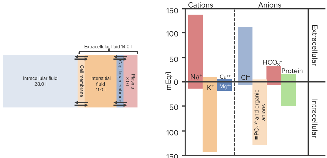
#Diagram body fluid compartments plus
In traditional Starling view, the filtration Jv is balanced by capillary absorption of tissue fluids, plus some lymphatic return. The work of Robert Hahn shows that we can reasonably approximate extracellular fluid kinetics in clinical practice to a two-compartment model, the central and tissue volumes. RSE and GM sees the body fluid compartments existing in a state of dynamic equilibrium, not as static buckets of fluid.


 0 kommentar(er)
